Home » Without Label » Anatomy Muscles Pelvis / What's The Score With The Pelvic Floor For Women | KB Physio - Ascending colon superior mesenteric vein superior mesenteric artery gonadal vessels linea semilunaris abdominal aorta linea alba inferior vena cava inferior mesenteric artery infe.
Anatomy Muscles Pelvis / What's The Score With The Pelvic Floor For Women | KB Physio - Ascending colon superior mesenteric vein superior mesenteric artery gonadal vessels linea semilunaris abdominal aorta linea alba inferior vena cava inferior mesenteric artery infe.
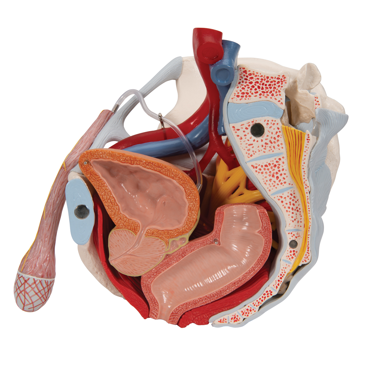
Anatomy Muscles Pelvis / What's The Score With The Pelvic Floor For Women | KB Physio - Ascending colon superior mesenteric vein superior mesenteric artery gonadal vessels linea semilunaris abdominal aorta linea alba inferior vena cava inferior mesenteric artery infe.. The root contains three erectile tissues (two crura and bulb of the penis), and two muscles (ischiocavernosus and bulbospongiosus). Muscles in the neck of bladder must contract and cause the internal urethral orifice to open before the detruser muscle can void the bladder: The muscles of the pelvic floor are collectively referred to as the levator ani and coccygeus muscles. Each hip bone, in turn, is firmly joined to the axial skeleton via its attachment to the sacrum of the vertebral column. These muscles, including the gluteus maximus and the hamstrings, extend the thigh at the hip in support of the body's weight and propulsion.
The bony pelvis consists of the two hip bones. The medial surface provides attachment for both transverse perinei, obturator internus and externus, piriformis, coccygeus and levator ani muscles. Each hip bone, in turn, is firmly joined to the axial skeleton via its attachment to the sacrum of the vertebral column. These muscles move the thigh toward the body's midline. These muscles have attachments to the pelvis as follows:
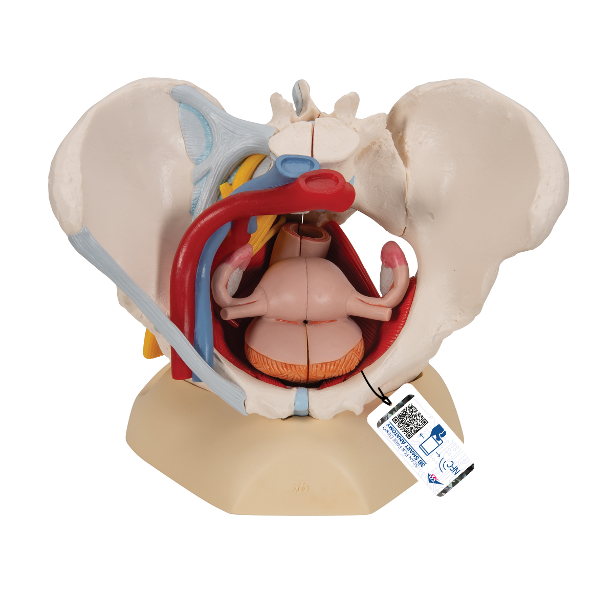
Male Pelvis Skeleton Model with Ligaments, Vessels, Nerves ... from www.3bscientific.co.uk Included in this group are the adductor longus, adductor brevis, adductor magnus, pectineus, and gracilis muscles. These muscles have attachments to the pelvis as follows: The medial surface provides attachment for both transverse perinei, obturator internus and externus, piriformis, coccygeus and levator ani muscles. (2) the levator ani and the coccygeus, which together form the pelvic diaphragm and are associated with the pelvic viscera. The function of the pelvic floor is to help assist with child birth, prevent incontinence and support organs within the pelvis. The muscles within the pelvis may be divided into two groups: Large ligaments, tendons, and muscles around the hip joint hold the bones (ball and socket) in place and keep it from dislocating. The floor of the pelvis is made up of the muscles of the pelvis, which support its.
Attached to the pelvis are muscles of the buttocks, the lower back, and the thighs.
The muscles of the penis include the corpora cavernosa,. Area between the asis (anterior superior iliac spine) and aiis (anterior inferior iliac spine). (2) the levator ani and the coccygeus, which together form the pelvic diaphragm and are associated with the pelvic viscera. Structure of the pelvic girdle. It connects the axial skeleton to the lower limbs. Muscles in the neck of bladder must contract and cause the internal urethral orifice to open before the detruser muscle can void the bladder: Clinical anatomy the lumbosacral trunk (l4, l5) and the ventral ramus of nerve s1 cross the nerves of the pelvis surface of the joint and may be involved in the disease of the joints, causing pain in the area of their distribution below the knee. The main function of the pelvic floor muscles are: The pelvis's frame is made up of the bones of the pelvis, which connect the axial skeleton to the femurs, and therefore acts in weight bearing of the upper body. It provides attachment to some important muscles in the region, and forms a cavity which accommodates several important internal organs. The levator ani muscles are the largest group of muscles in the pelvis. Piriformis the piriformis is a triangular muscle 1 on either side on the very front of the posterior wall of true pelvis. It originates from the pelvic outermost layer of the middle 3 sections of sacrum by 3 digitations.
The main function of the pelvic floor muscles are: Structure of the pelvic girdle. The muscles of the pelvic floor are collectively referred to as the levator ani and coccygeus muscles. Piriformis the piriformis is a triangular muscle 1 on either side on the very front of the posterior wall of true pelvis. Clinical anatomy the lumbosacral trunk (l4, l5) and the ventral ramus of nerve s1 cross the nerves of the pelvis surface of the joint and may be involved in the disease of the joints, causing pain in the area of their distribution below the knee.
Anatomical Teaching Models | Plastic Human Pelvic Models ... from www.a3bs.com It is located in the superficial perineal pouch of the pelvic floor, and is not visible externally. They form a large sheet of skeletal muscle that is thicker in some areas than in others. Muscles an important group of muscles in the pelvis is the pelvic floor. Muscles in the neck of bladder must contract and cause the internal urethral orifice to open before the detruser muscle can void the bladder: Psoas consists of a pair of deep muscles (psoas major and iliacus) located on each side of the pelvis in the abdomen. This mri male pelvis axial cross sectional anatomy tool is absolutely free to use. The penis can be anatomically divided into three parts: From there, it travels through the rectum and out of the anus.
The pelvis is a basin shaped bony structure formed by the combination of two pelvic bones (hip bones or innominate bones) and the sacrum.
On the posterior side they are the glutei and on the anterior side the hip muscles extending into the thighs. The levator ani is a broad sheet of muscle. Clinical anatomy the lumbosacral trunk (l4, l5) and the ventral ramus of nerve s1 cross the nerves of the pelvis surface of the joint and may be involved in the disease of the joints, causing pain in the area of their distribution below the knee. The function of the pelvic floor is to help assist with child birth, prevent incontinence and support organs within the pelvis. It provides attachment to some important muscles in the region, and forms a cavity which accommodates several important internal organs. Structure of the pelvic girdle. The main function of the pelvic floor muscles are: The penis can be anatomically divided into three parts: These muscles, including the gluteus maximus and the hamstrings, extend the thigh at the hip in support of the body's weight and propulsion. It attaches to the walls of the lesser pelvis, separating the in this article, we shall look at the anatomy of the muscles that make up the inferior lining of the cavity; The muscles within the pelvis may be divided into two groups: It's supplied by ventral rami of first and 2nd sacral nerves (s1, s2). Ascending colon superior mesenteric vein superior mesenteric artery gonadal vessels linea semilunaris abdominal aorta linea alba inferior vena cava inferior mesenteric artery infe.
It provides attachment to some important muscles in the region, and forms a cavity which accommodates several important internal organs. The pelvis also houses the reproductive organs, which have their own muscles. These muscles, including the gluteus maximus and the hamstrings, extend the thigh at the hip in support of the body's weight and propulsion. Psoas consists of a pair of deep muscles (psoas major and iliacus) located on each side of the pelvis in the abdomen. These muscles have attachments to the pelvis as follows:
Muscles Of Pelvis Floor Cross Section Photograph by ... from render.fineartamerica.com The penis can be anatomically divided into three parts: The medial surface provides attachment for both transverse perinei, obturator internus and externus, piriformis, coccygeus and levator ani muscles. Muscles in the neck of bladder must contract and cause the internal urethral orifice to open before the detruser muscle can void the bladder: It provides attachment to some important muscles in the region, and forms a cavity which accommodates several important internal organs. Each hip bone, in turn, is firmly joined to the axial skeleton via its attachment to the sacrum of the vertebral column. These muscles have attachments to the pelvis as follows: The pelvis also houses the reproductive organs, which have their own muscles. The right and left hip bones also converge anteriorly to attach to each other.
The penis can be anatomically divided into three parts:
Structure of the pelvic girdle. The pelvic floor muscles include; It is located in the superficial perineal pouch of the pelvic floor, and is not visible externally. Area between the asis (anterior superior iliac spine) and aiis (anterior inferior iliac spine). Arcus tendineus levator ani and the ischial spine This mri male pelvis axial cross sectional anatomy tool is absolutely free to use. Piriformis the piriformis is a triangular muscle 1 on either side on the very front of the posterior wall of true pelvis. The pelvic cavity and perineum. The medial surface provides attachment for both transverse perinei, obturator internus and externus, piriformis, coccygeus and levator ani muscles. It is strengthened and supported by several joints and ligaments. It can be described as one of the bodies diaphragms. Ƒ pelvic floor dysfunction is common and. The right and left hip bones also converge anteriorly to attach to each other.